Category: Articles
Torn meniscus? Faster Recovery, Better Knees: The Benefits of Meniscus Preservation
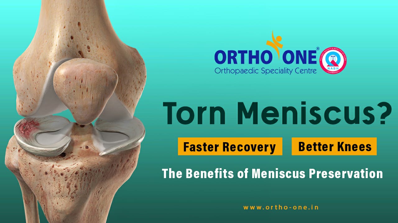
Those precious knee cushions, the menisci, play a vital role in shock absorption and joint stability. But a twist, a jump, or simply wear and tear can lead to tears, causing pain, swelling, and hindering your active lifestyle. Ouch!
Traditionally, a torn meniscus meant partial removal, potentially leading to early knee Arthritis. But here’s the good news: Meniscus Preservation Surgery is changing the game!
But what exactly is Meniscus Preservation, and why should you consider it?
Dr. David V Rajan, a renowned orthopaedic surgeon at Ortho One, Coimbatore, sheds light on this crucial procedure. He emphasizes that the meniscus acts as a shock absorber and stabilizer, preventing excessive wear and tear on the joint. When tears occur, traditional surgery often involves partial or complete removal, which can lead to early arthritis and decreased function.
However, meniscus preservation techniques focus on repairing or stitching the torn meniscus, allowing it to heal and regain its vital role. This minimally invasive approach boasts several advantages:
1. Faster recovery and rehabilitation: Say goodbye to lengthy post-surgery woes! Meniscus preservation typically translates to quicker healing times and a smoother return to your active lifestyle.
2. Reduced risk of Arthritis: Preserving the meniscus helps maintain the natural mechanics of the knee joint, significantly lowering the risk of developing arthritis later in life.
3. Improved joint function and stability: A healthy meniscus means a happier knee. You’ll experience better stability, reduced pain, and a wider range of motion, letting you conquer those stairs and slopes with confidence.
Dr. David V Rajan, a pioneer in meniscus preservation techniques, utilizes advanced arthroscopic procedures to achieve optimal outcomes. His expertise and commitment to minimally invasive approaches make him a trusted choice for patients seeking to save their menisci and safeguard their long-term knee health.
According to Dr. David V Rajan, “ An early diagnosis is key to a faster recovery, better joint stability, and long-term benefits for active individuals.”
So, if a torn meniscus has you sidelined, don’t settle for outdated solutions! Head to Ortho One and let Dr. David V Rajan explore meniscus preservation for you.
Remember:
1. Not all meniscus tears require surgery.
2. Meniscus preservation is a viable option for many patients.
3. Early intervention is key for successful outcomes.
Do your research, ask questions, and choose an orthopaedic surgeon who prioritizes your long-term joint health.
Navigating the Silent Epidemic: Understanding the Link Between Obesity and Early Arthritis
In recent years, a troubling shift has occurred in the landscape of health concerns. Obesity, once primarily associated with older generations, has now permeated the lives of young people, posing a significant threat to their well-being. Dr. P. Yuvarajan of Ortho One sheds light on this worrisome trend, attributing it to the Westernized embrace of sedentary lifestyles, stress-induced unhealthy eating, and a lack of exercise. The repercussions are profound, with a surge in Body Mass Index (BMI) emerging as a direct contributor to knee-joint pain and the onset of early arthritis.
The Impact of Excess Weight on Knee Health:
Excess weight is not merely a cosmetic concern; it acts as a relentless force that can have detrimental effects on the delicate structures surrounding the knee. Dr. Yuvarajan vividly describes this impact as a constant barrage of pressure, gradually wearing down the protective cushion of cartilage and soft tissues. The result is a breakdown that, when coupled with inflammation triggered by obesity, creates a fertile ground for the development of early arthritis. What was once considered an affliction of seniors has now become a looming threat to the mobility and well-being of the younger generation.
Breaking Down the Mechanics: Obesity as a Bully to Your Knees:
To understand the gravity of the situation, envision excess weight as a bully that pummels the knee’s cartilage and soft tissues. Dr. Yuvarajan’s insights emphasize how this relentless force leads to premature wear and tear, akin to a fragile cushion constantly under attack. The imagery paints a stark picture of the toll obesity takes on the knees, ultimately paving the way for the early onset of arthritis.
The Wake-Up Call:
The urgency to address this silent epidemic is clear. It’s time to break free from the unhealthy habits that fuel obesity and its subsequent impact on joint health. Dr. Yuvarajan advocates a three-pronged approach: prioritize movement, embrace a balanced diet, and seek guidance from experts in the field. This wake-up call is an invitation to take control of your health and, in doing so, protect not only your knees but also secure a pain-free future.
Choosing a Healthier Path:
For young individuals, the shadow of obesity looms large, driven by a toxic mix of stress, unhealthy food choices, and a sedentary lifestyle. Dr. Yuvarajan’s expertise highlights the pivotal role that a high BMI plays in early knee-joint pain and arthritis. The call to action is clear: step up, ditch the couch, and choose a healthier path. By doing so, you can safeguard your knees, reclaim the joy of movement in the prime of life, and embrace a future free from the shackles of pain.
Conclusion:
Understanding the intricate link between obesity and early arthritis is the first step towards a healthier future. Dr. P. Yuvarajan’s insights serve as a guiding beacon, urging us to break free from the silent epidemic and make informed choices that prioritize our well-being. It’s time to protect our knees, embrace movement, and pave the way for a pain-free journey through life.
Beyond Meniscectomy The Crucial Role of Meniscal Preservation in Knee Health
The meniscus is a cartilage present in the knee that acts as a cushion or shock absorber. Each knee has two menisci which are very important for the stability of the knee. Meniscus tears can happen due to trauma or because of wear and tear in the aging population. Initially, meniscectomy was the most common procedure followed to treat meniscal tears, where portions of the damaged meniscus were either partially or completely removed. However, recently there has been a debate going on whether to excise the meniscus or preserve it during knee surgeries. Dr. David V Rajan, a world-renowned Arthroscopic knee and shoulder surgeon, answers this important question and discusses the reasons for his opinion.
“Do you think that the shock absorbers in your two-wheelers or four-wheelers are essential for a smooth ride? Any mechanic would let you know that without them our vehicles will get completely damaged. Similarly, the knee meniscus, which is the shock absorber of our knees, is essential for a healthy knee joint. If there is any damage to the meniscus, it is essential to repair or restore it, without which the knee joint will be damaged due to wear and tear and will end in osteoarthritis,” says Dr. David V Rajan.
“Any patient who undergoes ACL surgery or meniscus surgery under my care can rest assured that we will do our best to preserve the meniscus as much as possible,” assures Dr. David V Rajan. There are many different techniques that can be used to achieve this including passing sutures, using implants, and all-inside, inside-out, outside-in. All these techniques can be used together as a combination or one of these techniques can be used depending on the extent, location, and severity of the meniscal tear.
Many people can continue with their normal routine without much discomfort during the initial few days after a meniscal tear happens. Due to this, there is a delay in consulting a doctor under the misconception that the injury is minor and will self-heal. According to Dr. David V Rajan, “Repairing the meniscal tear can be done very easily if the patient approaches the doctor at an early stage. However, the more he/she delays getting treatment meniscal repair and preservation gets harder to achieve.” Hence, it is very important to consult a doctor immediately if you have had a fall or if your knee feels swollen, tender, or unstable.
After meniscal repair surgery, the patient will have to go non-weight bearing or have a prolonged rehabilitation period. This will help the meniscus to heal completely allowing complete recovery. However, Dr. David V Rajan believes the delays and difficulties faced during the recovery period are a small price to pay for knee health which the patient can enjoy for a very long time. When the meniscus is repaired well or preserved efficiently, the patient can continue to participate in sports or partake in any activity of their choice without any discomfort or pain.
Dr. David V Rajan is therefore of the opinion that meniscal preservation is absolutely mandatory and has to be done whenever possible. Maintaining your shock absorbers will help the knee joints to function without any pain and plays a vital role in maintaining a healthy and active lifestyle.
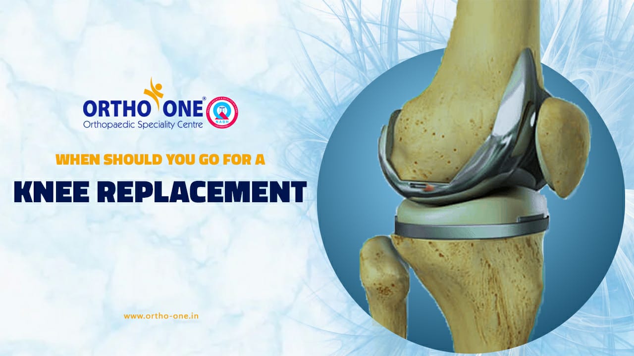
Knee pain can be the most frustrating condition that can hamper one from leading a normal life. Such conditions require a very cautious and tailored approach. There are many surgical and non-surgical ways to treat a knee injury, but there comes a time when a knee replacement becomes the best option for regaining mobility which can aid in improving the quality of life. However, understanding when to go for a knee replacement is a very important decision that needs to be made.
What is Knee Replacement Surgery?
Through knee replacement surgery, damaged or worn-out knee joints are replaced in part. The procedure aids in improving knee function and reduces discomfort. Damaged bone and cartilage are rebuilt during the procedure with metal and plastic components.
It is important to know that a knee replacement is not an option for early knee pain. A surgeon will be in a better position to tell if a knee replacement is needed or not. The surgeon examines your knee’s strength, stability, and range of motion in order to get a better understanding of the condition. X-rays are used to assist determine the level of damage.
From the Doctor’s Desk on Knee Pain and Knee Replacement Surgery
Dr. P. Yuvarajan, an expert at Ortho One Orthopedic Speciality Center, sheds light on knee pain and when the need for surgical intervention arises. The knee joint is a complex structure that constitutes both bone and soft tissue. Dr. Yuvarajan emphasizes that soft tissue pathologies in patients can often be managed without surgery. These pathologies may involve issues with the meniscus, ligaments, collateral ligaments, or even hip joint pain. In such cases, non-surgical treatments are advised, such as medication and physiotherapy.
However, if the pain persists despite conservative therapy, further evaluation is necessary to identify the root cause of the knee pain. Even if the specific reason for the pain is apparent, patients need to understand that knee pain won’t magically disappear with medication alone. Regular follow-ups are a must so that we can monitor the progress.
During the initial consultation with a doctor, typically, an X-ray and blood investigations are conducted to determine the cause of the knee pain. In the first session, patients are usually prescribed medications and physiotherapy. Then follow-up appointments, scheduled at intervals of 2 to 3 weeks depending on the patient’s needs, allow the doctor to assess progress.
If, in the second follow-up, the pain has completely subsided, patients can continue with medical treatment and physiotherapy. However, if pain persists despite the treatments further investigation would be conducted to identify the source of the pain. Even when the knee joint’s cartilage appears normal in tests, an MRI can reveal tears or damage to the joint. In such cases, minimally invasive arthroscopic procedures are conducted to address the issues.
It’s important to bust the myth that all cases of knee pain require surgery. In reality, approximately 70% of patients with early knee pain present with issues related to soft tissue rather than cartilage problems. These patients are initially managed non-surgically. The decision to proceed with surgery is made based on various factors, like the presence of deformities or significant bone changes seen in X-rays.
Advanced arthritis, characterized by a lack of space between the thigh bone and leg bone, may indicate the need for surgical procedures, such as partial or total knee replacement. Thus, the decision for surgery is based on a combination of clinical, radiological, and symptomatic criteria.
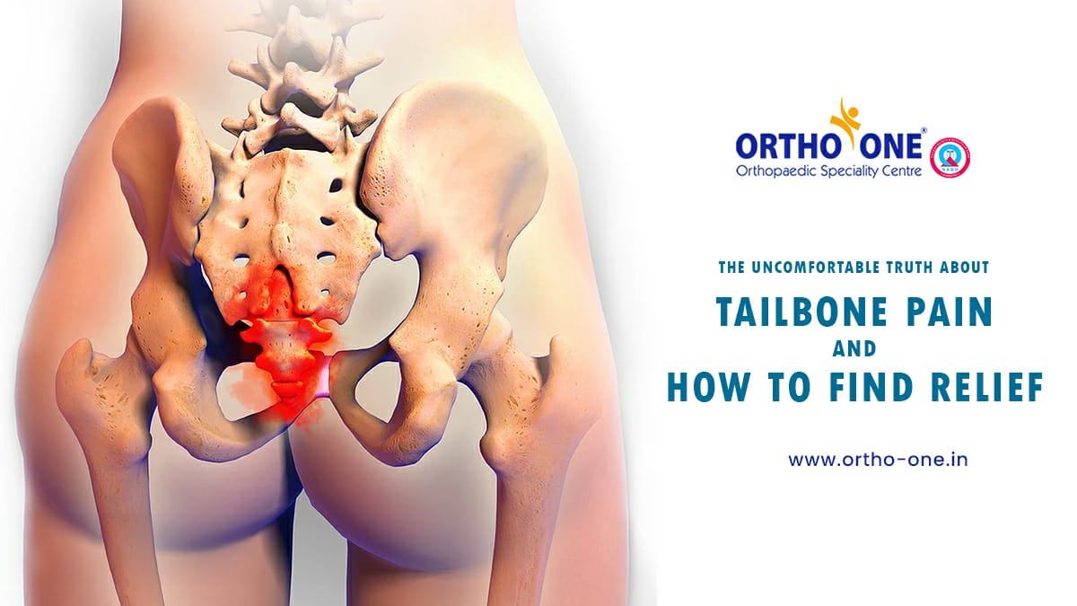
Dr. R. SreeRamaligam, spine surgeon at Ortho-One in Coimbatore, discusses tailbone pain, its causes, and treatment options. Tailbone pain is commonly experienced by people who participate in activities that require prolonged sitting, such as bike rides or long-distance travel. Trauma due to adventure sports can also lead to tailbone pain. Such activities can put pressure on the tailbone, causing changes that can lead to tailbone pain. This is known as coccydynia or coccygeal pain in medical terms.
“The tailbone is the backbone’s final and lowest segment, and it can move independently. As a result, there are numerous ways for the tailbone to get damaged or change position causing pain,” Dr. R. SreeRamalingam says. Some people experience this change as a bone subluxation, in which the tailbone moves up. In some cases, the tailbone can become dislocated and completely removed from its original location. It could be a simple sprain in others, or it could become lodged in the upper bone of the spinal cord. All of these can result in tailbone pain or pain in the area where the tailbone touches the surface while sitting.
“The easiest way to deal with tailbone pain is to sit in such a way that there is no pressure on the tailbone. This entails leaning forward while sitting down by sitting on a donut-shaped pillow,” recommends Dr. R. SreeRamalingam.
In the early stages, tailbone pain has a good chance of healing on its own if precautions are taken to avoid putting pressure on the tailbone for at least 2-3 weeks. Painkillers can be taken, if necessary, during this self-care phase. Dr. R. Sreeramalingam, however, advises against using over-the-counter ointments. This is due to the fact that most analgesic ointments contain ingredients that cause a burning sensation. As a result, applying such ointments in the tailbone area, which is very close to the anal region, can cause burning in the sensitive anus. Therefore he recommends only adequate rest and painkillers and not analgesic creams or ointments.
“Even After adequate resting for 3 weeks if the pain refuses to subside, then it is time to visit a doctor,” says Dr. R. SreeRamalingam. The doctor will use special X-rays called Coccyx view to view the position of the tailbone and to check if it is aligned correctly or if it has been pushed to the top or bottom. After finding out the changes, if any, to the tailbone, treatment must be designed accordingly.
Most people find that exercises and ointments designed specifically for tailbone pain work wonders. However, if these measures do not relieve the tailbone pain, Dr. R. SreeRamalingam recommends a coccyx block injection. This is administered in the joint of the tailbone after placing the patient in a facedown position. Following the injection, the tailbone is repositioned. This gives immediate pain relief, allowing the patient to sit on hard surfaces and carry on with their day-to-day activities.
“There are no surgical interventions that can be used to treat the tailbone pain. The coccyx block acts as a final and very effective method to treat tailbone pain,” assures Dr. R. SreeRamalingam.
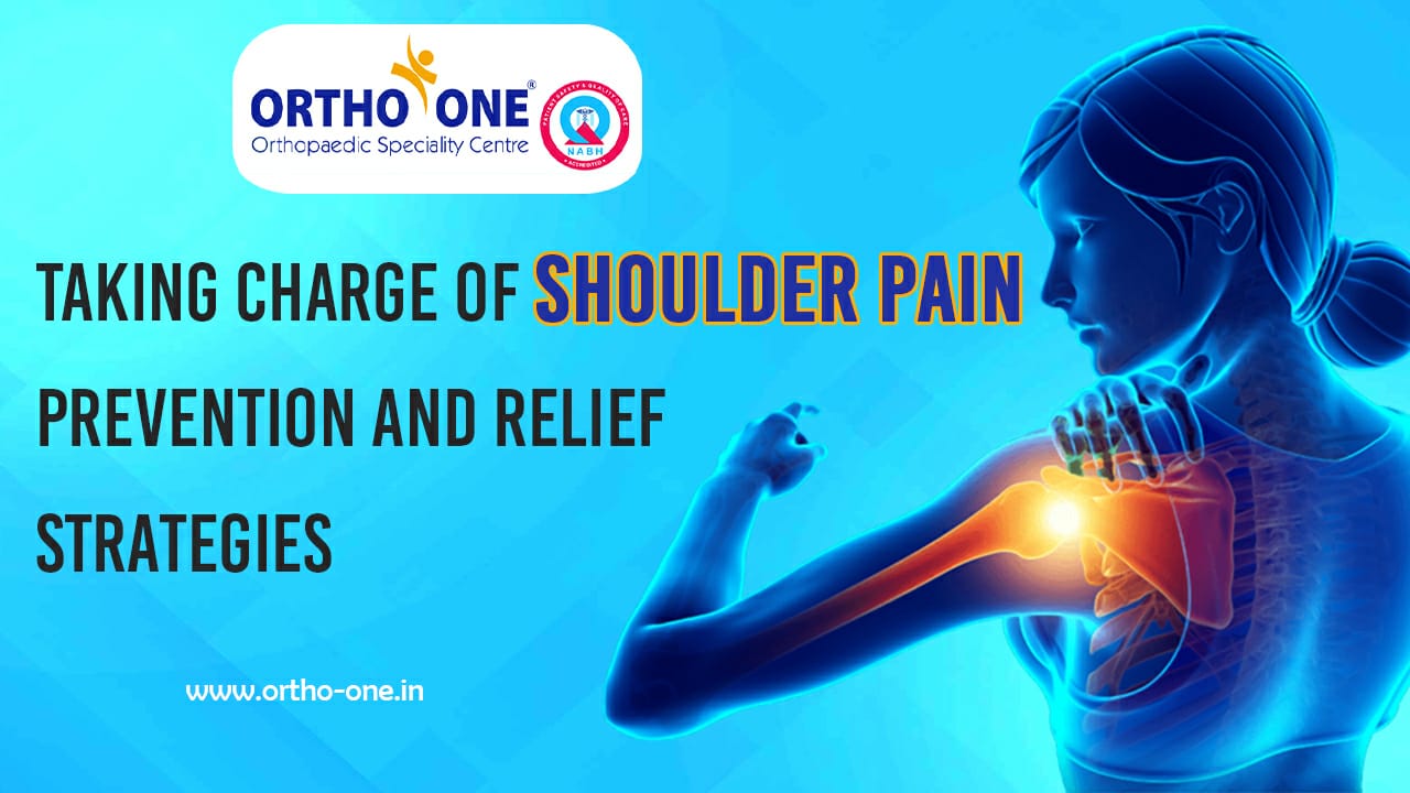
‘How to avoid or prevent shoulder pain?’ is a question that is often directed at Dr. M Shyam Sundar, shoulder and elbow specialist, Ortho-One, Coimbatore. “While it may not be possible to avoid shoulder pain, there are steps that can be taken to address the problem once it occurs,” Dr. Shyam responds.
Shoulders are very complex joints made of multiple bones, muscles, and tendons. This ball and socket joint allows for a variety of shoulder movements that we use in our daily lives, such as flexion, extension, external rotation, and internal rotation. The shoulder joint’s versatile movements make it vulnerable to a variety of problems that cause shoulder pain. “Shoulder pain is a very common problem that people of all ages face, and understanding how it occurs can help us prepare and take steps to avoid it,” says Dr. Shyam.
There are multiple reasons for the occurrence of shoulder pain. Young people may experience shoulder pain as a result of accidents or overuse injuries. Overuse injuries are those caused by repetitive actions that cause muscle overactivity, resulting in fatigue and pain. This is very commonly noted in athletes participating in overhead sports such as swimming, tennis, and volleyball. Another common occurrence among athletes is scapular malposition, which occurs when the shoulder bone in the back does not function properly, thereby interfering with normal shoulder movements. Scapular malpositions are classified into different types based on the muscles that are affected and the type of movement it impedes. Among older people, shoulder impingement is the most common cause of shoulder pain, which occurs when the bone pinches the tendon while lifting the hand, causing pain.
“It is impossible to prevent shoulder pain in either of these age groups, but we can learn how to manage it if it occurs,” says Dr. Shyam. RICE is the recommended treatment for shoulder pain caused by impingement or overuse syndrome. RICE is an acronym that stands for rest, ice application, compression, and elevation. Dr. Shyam believes that the most important factor in dealing with shoulder pain is rest. The best way to reduce shoulder pain is to get plenty of rest and avoid the movements that cause it. The next most important step is to pinpoint the specific movement that is causing the shoulder pain and perform corrective exercises tailored to that movement. Athletes can avoid scapular malposition by improving their posture and learning proper shoulder conditioning exercises. These exercises can also be adopted by older people who have shoulder pain in order to improve their shoulder stability and movements.
“Shoulder pain can affect anyone at any age, regardless of activity level. However, it is possible to relieve shoulder pain with proper measures such as RICE therapy, corrective and/or shoulder conditioning exercises, maintaining proper posture, and so on. Therefore, it is important to make ourselves aware of the underlying cause for the shoulder pain and to seek appropriate treatment immediately,” says Dr. Shyam.
ஆர்த்ரிட்டிஸ் என்றால் என்ன என்ற சந்தேகம் உங்களுக்கு உள்ளதா? நீங்கள் முதலில் தெரிந்து கொள்ள வேண்டியது, ஆர்த்ரிட்டிஸ் (Arthiritis) அல்லது கீழ் வாதத்தால் அல்லது மூட்டுவலி என்பது ஒரு நோய் அல்ல; இந்த வார்த்தை மூட்டு வலி அல்லது மூட்டு நோயைக் குறிக்கிறது, மேலும் 100க்கும் மேற்பட்ட வகையான மூட்டுவலி மற்றும் தொடர்புடைய நிலைமைகள் உள்ளன. அனைத்து வயது, இனம் மற்றும் பாலின மக்கள் மூட்டுவலியுடன் வாழ்கின்றனர். இது வயதானவர்களைத் தாக்கும் நோய் அல்ல என்றாலும், சில வகையான மூட்டுவலி இளையவர்களை விட வயதானவர்களுக்கு ஏற்படுகிறது.
பொதுவான மூட்டுவலி அறிகுறிகளில் வீக்கம் (Swelling) , வலி (Pain) , விறைப்பு (Stiffness) மற்றும் மூட்டுகளில் இயக்கம் குறைதல் (Difficulty in moving a joint) ஆகியவை அடங்கும். அறிகுறிகள் லேசானது முதல் கடுமையானது வரை மாறுபடும் மற்றும் வந்து வந்தும் போகலாம். சிலருக்குப் பல ஆண்டுகளாக ஒரே மாதிரியாக இருக்கலாம், ஆனால் சிலருக்கு அறிகுறிகள் முன்னேறலாம் மற்றும் காலப்போக்கில் மோசமடையலாம். கடுமையான மூட்டுவலியானது நாள்பட்ட வலி, அன்றாடச் செயல்பாடுகளைச் செய்வதில் சிரமம் மற்றும் நடைப்பயிற்சி மற்றும் படிக்கட்டுகளில் ஏறுதல் போன்றவற்றை வலியூட்டுவதாகவும், கடினமானதாகவும் மாற்றும்.
மூட்டுவலி நிரந்தர மூட்டு மாற்றங்களையும் ஏற்படுத்தும். குமிழ் விரல் மூட்டுகள் போன்ற இவை தெரியும், ஆனால் பெரும்பாலும் சேதத்தை எக்ஸ்ரேகள் (X-Ray) மட்டுமே காண முடியும். சில வகையான மூட்டுவலி இதயம், கண்கள், நுரையீரல், சிறுநீரகம் மற்றும் தோல் மற்றும் மூட்டுகளைப் பாதிக்கிறது.
அனைத்து வகையான மூட்டுவலிகளுக்கும் ஒரே காரணம் என ஒன்று இல்லை. கீல்வாதத்தின் வகை அல்லது வடிவத்தைப் பொறுத்து காரணம் அல்லது காரணங்கள் மாறுபடும், அவற்றில் சில முக்கியமான காரணங்கள் காயம், அசாதாரண வளர்சிதை மாற்றம், மரபணு பரம்பரை தாக்கம் , ஆர்.ஏ மற்றும் லூபஸை ஏற்படுத்தும் வகை போன்ற நோயெதிர்ப்பு அமைப்பு செயலிழப்பு ஆகும்.
பெரும்பாலான வகையான கீல்வாதம் காரணிகளின் பல்வேறு வகைகளாக இணைக்கப்பட்டுள்ளது. இருப்பினும், சிலருக்கு வெளிப்படையான காரணம் எதுவும் இல்லை மற்றும் அவற்றின் தோற்றத்தில் கணிக்க முடியாததாகத் தோன்றுகிறது.
ஆர்த்ரிட்டிஸால் அதிகம் பாதிக்கப்படுவது முழங்கால் முட்டு. இருப்பினும் ஆர்த்ரிட்டிஸ் என்பது பல்வேறு வகையான இன்னல்களை ஏற்படுத்தக்கூடிய ஒரு நிலை ஆகும். முட்டு வலியில் தொடங்கி உடம்பில் உள்ள இன்னல்கள் படிப்படியாக வளர்ந்து ஆர்த்ரிட்டிஸ் என்னும் நிலையை அடைகிறது. பெரும்பாலும் முழங்கால் முட்டு ஆர்த்ரிட்டிஸால் பாதிக்கப்படக் காரணம் உடலின் எடையைத் தாங்கும் பகுதி அது. நாம் நடக்கும்போதோ ஓடும்போதோ அல்லது வேறு எந்த ஒரு செயலையும் செய்யும் போதும், நமது உடலின் முழு எடையையும் தாங்குவது நமது முட்டு தான். அது மட்டும் அல்லாது நாம் செய்யும் செயலிற்குத் தேவையான சக்தியைப் பூமிக்குச் செலுத்துவதும் அதன் வேலை. இதனால் நாம் எந்தவொரு அன்றாட வேலை செய்யும் பொழுதும் அதற்குத் தேவையான பலத்தைத் தருவதோடு நாம் தரும் உராய்வுகளையும் (Friction) அது தாங்க வேண்டியது அவசியம். முழங்கால் முட்டு ஆர்த்ரிட்டிஸ் நாளடைவில் பல்வேறு சிரமங்களை ஏற்படுத்தும் என ஆய்வுகள் குறிப்பிடுகின்றன. ஒருவருக்கு முழங்கால் முட்டில் ஆர்த்ரிட்டிஸ் ஏற்பட்டால் காலப்போக்கில் நடப்பது ஓடுவது போன்ற அதிக பாதிப்பு ஏற்படும் செயல்களால் தேய்மானம் ஏற்படும். ஒருவரின் நிலைக்கு பொறுத்தவாறு அவர் செய்யும் செயலை பொருத்தும் அது ஏற்படுத்தும் பாதிப்பைப் பொருத்தும் எந்த தசைநார்களில் (Ligament) பாதிப்பு ஏற்பட்டு இருக்கிறது என்பதைப் பொருத்தும் ஆர்த்ரிட்டிஸின் பாதிப்பு நிலை ஏற்படுகிறது என்கிறார், மருத்துவர் பி. யுவராஜன், எம். எஸ். ஆர்த்தோ, ஆர்த்தோ ஒன் மருத்துவமனை.
When Devaraj, 74, began experiencing back pain, everyone in his family, including himself, blamed it on his age. “Anyone this old will obviously have back pain due to bone degeneration or spondylosis. He should probably ease up and change his lifestyle to be less active from now on,” they said.
Dr. Sreeramalingam, a spine surgeon at Ortho-One Orthopaedic Speciality Centre in Coimbatore, disagrees. There are numerous myths about back pain in the elderly. Many people believe that back pain is caused by old age. Back pain is also thought to be caused by spondylosis, or degenerative changes in the spine. “If degenerative changes caused by aging were the only cause of back pain, then every 70 or 80-year-old person would have back or neck pain. However, only a small percentage of the elderly suffer from severe back pain or spine-related pain,” says Dr. Sreeramalingam. Although some disability is to be expected with advancing age,
Dr. Sreeramalingam believes that not all pain experienced by the elderly is due to age-related degenerative changes.
The spine is made up of motion segments, which are the basic units that control movement. It is composed of two vertebral bodies separated by a disc and has two moving facet joints in the back. The disc degenerates as we age, causing the vertebral bodies to point outward, a condition known as Osteophyte formation. The vertebral bodies eventually settle into these new positions with very little disc in between. These changes in the motion segment are usually not painful. As a result, while all elderly people will experience these normal degenerative changes, not all will experience back pain.
There are four major underlying causes of back pain in the elderly. These include:
1. Disc problems in which the disc material protrude and press on the nerves.
2. Degenerative spondylolisthesis caused by micro-instabilities in the vertebrae, resulting in the vertebral bone slipping on top of the next vertebral bone during movement.
3. Lumbar canal stenosis, a condition in which the spinal canal through which the spinal cord travels becomes narrow and presses on the nerves.
4. Cancer metastasis or other diseases manifesting in the spine from elsewhere in the body.
“These underlying causes of back pain or neck pain are not similar across the age groups and therefore such back pains should not be ignored in the elderly,” says Dr.Sreeramalingam. The elderly face additional challenges as a result of degenerative changes in the body caused by aging, which must also be addressed. Back or neck pain in the elderly, according to Dr. Sreeramalingam, deserves the same attention as back pain in much younger people.
People like Devaraj require medical attention and an accurate diagnosis to determine the source of their back pain. “Treating the underlying condition often helps the elderly live a pain-free and fulfilling life,” says
Dr. Sreeramalingam. Rather than restricting or ignoring the elderly’s symptoms, or simply chalking it up to old age, families must ensure that the elderly with back pain see a doctor and receive a timely treatment.
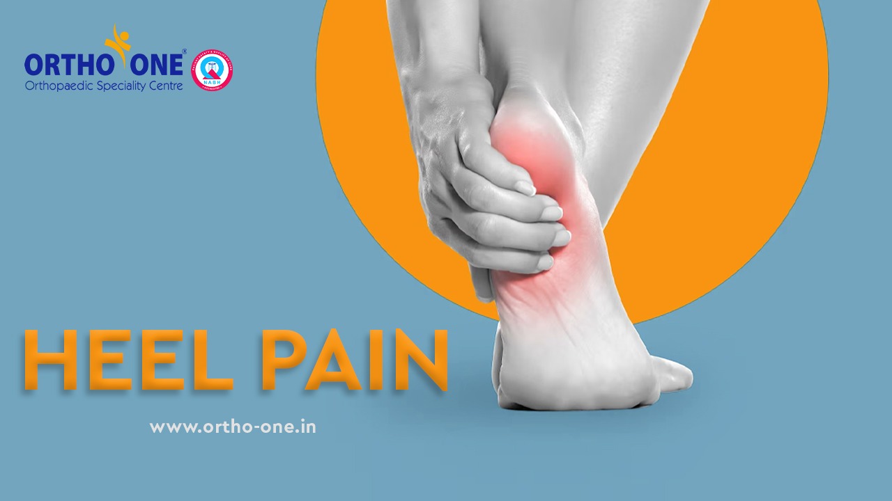
Do you dread getting out of bed in the morning because of a hurting foot? Do you have to tread carefully for the first few minutes after a night’s sleep due to extreme foot pain? Do your feet ache when you stand up after sitting for an hour or so to rest? According to Dr B Vidya Sagar, foot and ankle surgeon at Ortho-One Orthopaedic Speciality Centre, Coimbatore, if you answered yes, you must be suffering from heel pain.
According to Dr Vidya Sagar, approximately 25–30% of patients visiting Ortho-One’s foot and ankle clinic have heel pain. Plantar fasciitis is the correct term for the condition. Flatfoot, obesity, a high body mass index (BMI), or even thyroid dysfunction could be the root of the heel pain. However, in more than 50% of cases, there is no obvious cause.
People in their 40s and the middle-aged are typically the ones who experience this condition. The main reason is that people at this age start to get comfortable in their careers and daily routines and start to lose interest in physical activities like sports, working out, etc. This causes the tendinous portions (calf and foot muscles) to tighten. “The tendinous muscles should ideally be flexible. One should be able to stretch or flex them on command; if not, they ought to return to their resting position,” explains Dr Vidya Sagar.
In people suffering from heel pain, the plantar fascia (foot muscle) fails to stretch when one gets up after a long period of rest because it shortens at rest. Microtears brought on by a delay in stretching may lead to inflammation. This inflammation is what causes heel pain, and it can also lead to calcaneal spurs on the front or back of the heel.
There are many remedies to reduce heel pain in the initial stages which include taking painkillers, NSAIDs, and modification of footwear. However, according to Dr Vidya Sagar, the most effective treatment is stretching the foot and calf muscles. These exercises must be done at least twice a day continuously for 2-3 months to get visible results. If the patient is in such pain that he/she cannot do the stretching exercises, a local mild steroid injection will be administered. “Do not be alarmed by the word ‘steroid injections’,” says Dr Vidya Sagar. He guarantees that they have no systemic effects and only act locally at the site of the pain. The pain will significantly diminish within 2-3 days of receiving a steroid injection, and patients will be able to perform the advised exercises.
The last resort is to surgically stretch the Gastrocnemius and plantar fasciitis muscles if the patients still experience pain despite receiving conservative treatment. According to Dr Vidya Sagar, “This type of surgery is performed rarely because the majority of patients respond well to the conservative treatment alone.”
Dr Vidya Sagar cautions that while most patients will experience relief from heel pain through conservative management, it is important to also treat any accompanying symptoms. These include managing thyroid levels, addressing obesity issues, and treating flatfoot with medial arch supports if alignment problems are present. “Most patients will experience a drastic reduction in heel pain within 2-3 months when these issues are addressed and stretching exercises are done regularly,” says Dr Vidya Sagar.
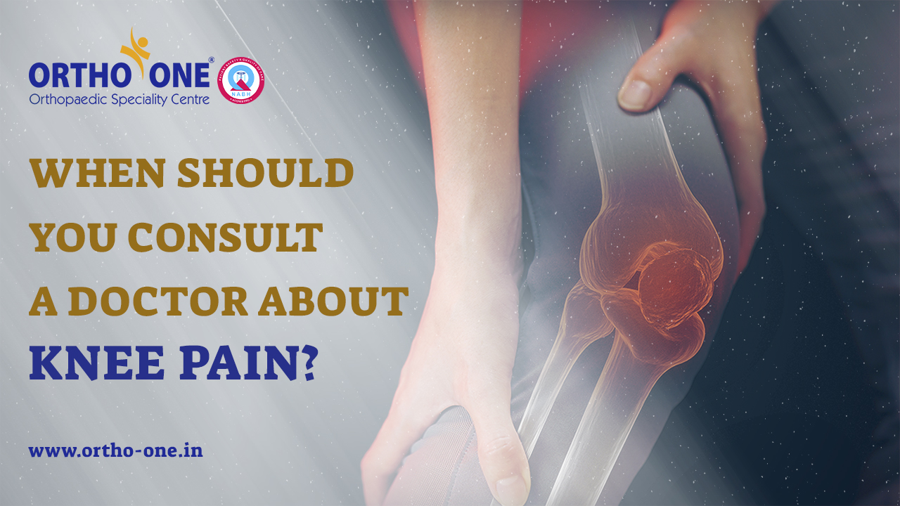
When should you consult a doctor about knee pain? How much pain should you be in before seeing a knee specialist? Dr Santosh Sahanand, a knee specialist at Ortho-One Orthopaedic Speciality Centre in Coimbatore, answers your burning questions and explains a few prevention strategies for knee pain.
The first point to consider is the intensity of the knee pain that you are experiencing. “Some people come to us as soon as the pain starts. Such patients will come even if the pain is very minor while there are some who refuse to meet a doctor even if they are in excruciating pain. It is always preferable to be somewhere in between these two extremes,” says Dr Santosh. If you are in a lot of pain and you are unable to put weight on your legs, then you must see a doctor. It is best to find the underlying cause of the pain and get treatment as soon as possible.
The second critical factor is the length of time you have been experiencing knee pain. “If you have insignificant pain, then you do not need to see a doctor. However, if this minor pain persists for a few days, it warrants a visit to the doctor,” says Dr Santosh. When detected in its early stages, the underlying problem causing the knee pain is usually smaller and can be easily treated. This is especially true for conditions such as joint degeneration caused by wear and tear. According to Dr. Santosh, there is a good chance of preventing knee joint degeneration even before it begins, but only if the problem is identified in its early stages.
“The most common mistake people make is assuming that knee pain occurs only when the knee joint wears out. What they do not realize is that knee pain can occur for a variety of other reasons too,” Dr Santosh says. The knee is a complex joint made up of many cartilage and bush-like tissues, and any bruises or injuries to any of these tissues can cause knee pain. When these injuries are minor, rest and certain exercises can heal the condition on their own. However, if one continues to play or put stress on the injured knees, the injury will get bigger and the pain will worsen rather than heal completely.
Therefore, it is always better to identify what is the underlying cause of your knee pain so that appropriate treatment can be initiated. “However, this does not mean that you should see your doctor for every minor ailment or pain,” Dr Santosh cautions. A knee pain that has been bothering you for more than a week or two or a knee pain that is becoming more intense, should be shown to a knee specialist, and thoroughly investigated.